Sarcomeres slide
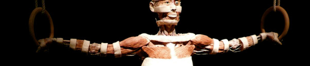
Skeletal muscle
The anatomical organisation of a skeletal muscle will be covered at its different levels, which are at the level of a/an:
The neuromuscular junction will also be discussed.
Skeletal Muscle
It is composed of an orderly arrangement of connective tissue and contractile cells. The epimysium is the external connective tissue wrapping around the entire muscle. The skeletal muscle is made up of fascicles.
Fascicle
Fascicles are bundles of individual muscle cells which make up a skeletal muscle as shown in Figure 1. These fascicles are surrounded by a connective tissue layer called the perimysium. There is a third connective tissue called the endomysium which separates and electrically insulates muscle cells from each other.

Figure 1. A skeletal muscle with associated connective tissues, showing structures all the way down to myofilaments.
Muscle Cell Fibre
Muscle cell fibres are so called because they are elongated. Its cell membrane is called the sarcolemma and the fibres have sarcoplasmic reticulum, which is its endoplasmic reticulum. T-tubules are the invagination of sarcolemma projecting deep down into the muscle cells. Sarcoplasmic reticulum form enlarged areas around T-tubules are called the terminal cisternae.

Figure 2. Structure of a skeletal muscle fibre containing myofibrils.
Myofibril
Myofibrils are the cylindrical bundles of contractile filaments. These individual contractile proteins are called myofilaments which are composed of thick and thin filaments. They form repeating units along the myofibril which are termed sarcomeres.
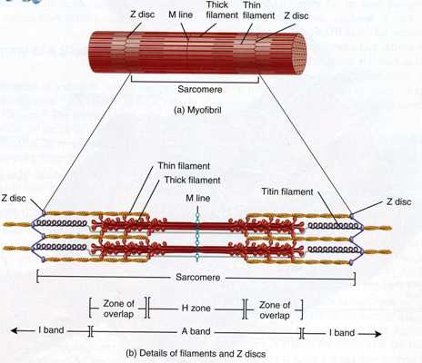
Figure 3. (a) Structure of a myofibril containing a sarcomere; (b) The overlapping pattern of thick and thin filaments.
Sarcomere
 The sarcomere is a functional contractile unit of a myofibril. It has:
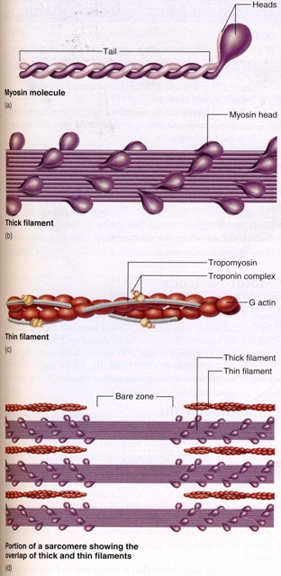
- Actin filaments - The thin filaments that consist of two intertwined chains of G actin molecules called troponin and tropomyosin. Tropomyosin is filamentous in structure and it coils around this actin filament. Troponin is a three part protein attached to the tropomyosin filaments. This is illustrated in Figure 4 to the right.
- Myosin filaments - The thick filaments that consist of 200 to 500 myosin molecules bundled together with the heads projecting outward in a spiral array. A single myosin molecule is composed of two intertwined polypeptides forming a filamentous tail and a double globular head, as shown in Figure 4.
Figure 4. Molecular structure of thick and thin filaments.
Actin and myosin filaments are abundant in skeletal and cardiac muscles which account for their striations. These striated muscles have dark A bands and lighter I bands as shown in Figure 5. The dark A-band has two parts. The darker area is where myosin filaments overlap actin filaments. H band is the lighter region in the middle of A band where the actin filaments do not reach. The I band consists of thin actin filaments anchored to Z-discs (composed of connectin proteins) bisecting through this I band. Sarcomere is the segment of myofibril from one Z-disc to the next one as shown in Figure 3. Z-discs are connected to the sarcolemma (muscle cell plasma membrane) by cytoskeleton. Individual sarcomeres shorten, bringing Z-discs closer to each other. The pulling of the sarcolemma achieves an overall shortening of a cell.

Figure 5. Five myofibrils of a single muscle fibre, showing the structure of sarcomeres and striation in relaxed state.
Neuromuscular junction
The neuromuscular junction is a synapse between a nerve fibre and a muscle fibre. Two neuronal cells are separated by a tiny gap called the synaptic cleft (about 60-100nm wide). A Schwann cell envelopes the entire neuromuscular junction, isolating it from surrounding tissue fluids. It prevents an electrical signal travelling down the nerve fibre from crossing the synaptic cleft; rather, it causes the nerve fibre to secrete a synaptic vesicle containing a neurotransmitter that stimulates the next cell.
Once an action potential arrives at the motor axon terminal, the membrane potential increases, activating voltage-gated Ca2+ channels. This allows Ca2+ ions to enter the axon terminal. Ca2+ ions make the synaptic vesicle fuse with the membrane of the axon terminal, releasing its neurotransmitter called acetylcholine at the neuromuscular junction. Acetylcholine depolarises the motor end plate by binding to acetylcholine receptors at this region. Na+ ions enter the cytosol and K+ ions leaves the cell. Acetylcholine is the only transmitter released at neuromuscular junction.
From the resulting depolarisation at the motor end plate, Ca2+ ions are released from the terminal cisternae into the cytosol. Ca2+ ions bind to troponin and tropomyosin that are present on actin filaments and trigger a contraction of the muscle. This process is illustrated in more detail in the "Sliding Filament Theory" section. After a brief time, the acetylcholine diffuses away from their receptor sites causing ion channels to close. The acetylcholine is then broken down by an enzyme called acetylcholinesterase present at the synaptic cleft, resulting in the relaxation of a muscle.

Figure 6. A neuromuscular junction.
References
- Figure 1 adapted from Tortora & Derrickson (2007, p. 293)
- Figure 3 adapted from Tortora & Derrickson (2007, p. 296)
- Figures 2, 4 and 5 adapted from Saladin (2003, p. 410-412)
- Figure 6 adapted from Saladin (2003, p. 415)